Monday 30 October 2023

Precision in Healthcare: Exploring ELISA Tests for HIV, HCV, Hepatitis B, and Syphilis Diagnosis by Trivitron Healthcare


purpose of elisa test

In the field of healthcare and diagnostics, precision is vital. Diagnostic tests are the cornerstone of accurate healthcare, and Trivitron Healthcare, a renowned name in the healthcare industry, recognizes the importance of precision. They have developed advanced ELISA (Enzyme Linked Immunosorbent Assay) test kits that excel in diagnosing critical infections such as HIV, HCV, Hepatitis B, and Syphilis with unwavering accuracy.


What is the ELISA Test Used For?


ELISA kits are instrumental in detecting and quantifying biological molecules like proteins, antibodies, hormones, and antigens, offering unparalleled sensitivity, specificity, and precision. These kits capture target molecules using immobilized capture antibodies and detect them with labeled antibodies, generating a signal that correlates with the molecule's concentration in the sample. ELISA technology has revolutionized disease diagnosis and research in healthcare.


How to Perform the ELISA Test?


Performing an ELISA test involves capturing the target molecule, typically a protein or antigen, using an immobilized capture antibody on a microplate. Then, a labeled antibody recognizing a different part of the target molecule is added. The label, often an enzyme, generates a signal, such as color change, fluorescence, or chemiluminescence, proportional to the target molecule's concentration. This process enables the accurate measurement of biomarkers, leading to disease diagnosis and monitoring drug therapies.


ELISA kits have numerous advantages over alternative methods, such as Western blot or radio-immunoassay. They are rapid, precise, and cost-effective, requiring only small sample sizes. They can also be automated for high-throughput screening. ELISA kits are versatile, offering a wide dynamic range of detection, from picograms to nanograms per milliliter.


Trivitron Healthcare's ELISA test kits are a testament to their commitment to precision. They have designed kits for diagnosing HIV, HCV, Hepatitis B, and Syphilis, each tailored to address specific health challenges. With their high sensitivity and specificity, these kits play a vital role in disease diagnosis, contributing to advancements in healthcare practices and medical research. Trivitron Healthcare's ELISA kits are more than a promise of precision; they are a reality that enhances patient lives and empowers the medical community.


Source

Friday 27 October 2023

Why Lead is the Best Protection from Radiation


In the field of medical technology, it is imperative to ensure that the radiation emitted by devices is appropriately contained to safeguard individuals from any harm to their bodies. The effects of radiation extend beyond immediate skin reactions like allergies, redness, burns, or hair loss. Yes, radiation can also harm cells by damaging their DNA, potentially impacting future generations as well. Consequently, the utilization of protective accessories such as radiation shields, aprons, gloves, and eyewear becomes crucial to mitigate these risks.


Medical professionals who perform interventional procedures face a higher risk of radiation exposure and should prioritize protecting their bodies by wearing radiation protection aprons, gloves, thyroid collars, protective headwear, and glasses specifically designed for radiation protection. Trivitron Healthcare offers radiation protection aprons designed to safeguard healthcare practitioners from the radiation emitted by medical imaging devices such as X-ray fluoroscopy during surgical procedures or diagnostic tests.


The Importance of Radiation Protection Aprons


Radiation protection aprons have become a vital component in the arsenal of tools that medical professionals use to shield themselves from the harmful effects of radiation. Among various materials used for these aprons, lead has emerged as the best choice for protecting against radiation. Here's why lead aprons are considered the gold standard in radiation protection:


1. Unparalleled Shielding Ability: Lead is a dense material that excels at blocking radiation. When X-rays or other forms of ionizing radiation are generated, lead aprons act as a formidable barrier. They effectively absorb and deflect the harmful rays, ensuring that they do not penetrate the wearer's body.


2. Durability: Lead aprons are known for their durability. When properly cared for, they can provide long-term protection. This is crucial, especially in the medical field, where aprons are frequently used and exposed to potentially damaging conditions.


3. Flexibility and Comfort: Lead aprons are designed with a focus on comfort. They are typically manufactured to be flexible, allowing medical professionals to move freely while maintaining their protective barrier. This flexibility ensures that the aprons can be worn for extended periods without causing discomfort.


4. Easy to Clean and Maintain: As mentioned in a blog from Trivitron Healthcare, regular maintenance is essential for the longevity of lead aprons. Proper cleaning and sanitizing are vital to prevent cracks or vulnerabilities in the apron material. The inspection process should include examining seams, covers, and belts for any signs of damage. This upkeep ensures that the apron maintains its structural integrity.


5. Responsible Disposal: When a lead apron is no longer safe for use, it's essential to follow proper disposal procedures to protect the environment. Lead is a hazardous material and should not be discarded in regular garbage. Contacting recycling companies that specialize in lead materials or local hazardous waste carriers for proper disposal is crucial.


Conclusion


In the world of radiation protection, lead aprons have established themselves as the best choice for safeguarding medical professionals and patients from the harmful effects of radiation exposure. Their unmatched shielding ability, durability, comfort, and ease of maintenance make them the preferred option in the medical field.


Proper care and maintenance of lead aprons, as outlined by Trivitron Healthcare, ensure that these essential protective garments remain effective over time. Furthermore, responsible disposal of damaged lead aprons is critical to protect the environment and adhere to safety regulations.


In summary, lead aprons are the gold standard for radiation protection in the medical industry, offering the best defense against the harmful effects of ionizing radiation. To learn more about the importance of clean radiation protection aprons, you can refer to Trivitron Healthcare's informative blog. For the best radiation protection accessories that meet industry needs, visit. Your safety and the well-being of future generations depend on it.

Wednesday 25 October 2023

Primary Antibody vs. Secondary Antibody in Clinical Diagnostics


primary antibody and secondary antibody

Antibodies are fundamental in both the human body and laboratory practices, particularly in immunoassays for disease detection. This article explores the distinctions between primary and secondary antibodies, their shared applications, and their roles in diagnostic tools, as discussed in the blog "Primary and Secondary Antibodies" by Trivitron Healthcare.


Primary antibodies are critical for identifying proteins and biomolecules in biological samples like blood, serum, and urine. They specifically bind to target antigens, aiding in the recognition of diseases such as cancer, diabetes, and neurological disorders. Notably, primary antibodies serve both diagnostic and therapeutic functions.


Secondary antibodies complement primary antibodies by binding to them, enhancing the detection of target proteins. These secondary antibodies come in different colors and offer increased sensitivity, making them suitable for applications like flow cytometry, western blotting, and cell imaging. They enable colorimetric, chemiluminescent, and fluorescent detection of primary antibodies, enhancing sensitivity and flexibility in visualizing target molecules.


Both primary and secondary antibodies are widely used in laboratory techniques, including western blotting, immunostaining, and ELISA. ELISA, in particular, employs capture antibodies, detection antibodies, and secondary antibodies. Primary antibodies form a "sandwich complex" by binding to target antigens, and secondary antibodies, if used, amplify the signal by recognizing and binding to the detection antibodies. This multi-step approach enables sensitive and specific antigen detection and quantification.


Trivitron Healthcare's ELISA kits, such as the HCV ELISA Test Kit, the HIV Ab/Ag ELISA Test Kit, and the Syphilis Ab ELISA Test Kit, exemplify the significance of primary and secondary antibodies in clinical diagnostics. These kits play a vital role in detecting specific antibodies or antigens in human serum or plasma, contributing to disease diagnosis and healthcare precision.


In conclusion, primary and secondary antibodies are indispensable components of clinical diagnostics. They facilitate accurate target identification, improving patient care and advancing medical research. Their critical roles are exemplified in ELISA kits, highlighting their importance in addressing health challenges and promoting healthcare advancements.


Source

Thursday 19 October 2023

Demystifying Dengue: How the Virus Attacks and Spreads


how does dengue attack the body

Dengue fever, a viral illness transmitted by Aedes mosquitoes, is a global concern, particularly in tropical regions. Understanding how dengue fever attacks the body and spreads is crucial for its management.


How Does Dengue Fever Attack the Body?

When an infected mosquito bites, the virus enters the body and triggers the immune system. It has two main components: the innate immune system offers immediate, non-specific protection, and the adaptive immune system provides specific, long-lasting defense. The virus infects skin and immune cells, including Langerhans cells. It then spreads to lymph nodes, bone marrow, spleen, liver, and blood, causing viremia. The adaptive immune response produces antibodies and cytotoxic T cells to combat the virus.


How Does Dengue Virus Evade the Immune System?

The virus infects monocytes and macrophages instead of being destroyed by them. These cells spread the virus via the lymphatic system, allowing it to persist and cause widespread infection.


Factors Contributing to Severe Dengue Infections

Chronic diseases and genetic variations can increase the risk of severe dengue infections.


Ways to Protect From Dengue

Preventing dengue involves eliminating stagnant water, using mosquito nets, wearing protective clothing, and applying mosquito repellents.


How is Dengue Virus Detected in the Human Body?

The NS1 antigen is vital for diagnosis, helping healthcare professionals confirm dengue infection quickly, enabling timely treatment.


In conclusion, understanding how dengue fever attacks the body is crucial for effective diagnosis and management. While the immune system plays a vital role, the virus has strategies to evade it. Ongoing research aims to enhance our knowledge of dengue fever and develop better prevention and treatment methods.


Source

Tuesday 17 October 2023

Decoding the Chest Pain Dilemma: Heartburn or Heart Attack?


heartburn or heart attack

Heartburn and heart attack are two terms often used interchangeably, but they signify vastly different medical issues. This article aims to clarify the differences between heartburn and a heart attack and help readers understand when they should seek medical attention.


Understanding Heartburn:

Heartburn occurs when stomach acid flows back into the esophagus, causing a burning sensation in the chest. It is typically triggered by consuming spicy or fatty foods, smoking, obesity, pregnancy, stress, or certain medications. Heartburn symptoms include an acidic taste in the mouth, regurgitation of food, and chest discomfort that worsens after meals. Over-the-counter antacids and lifestyle changes can alleviate heartburn.


Understanding a Heart Attack:

A heart attack is a life-threatening medical emergency that results from a blocked blood flow to the heart muscle, usually due to a blood clot. Symptoms of a heart attack can include chest pain or discomfort, pain radiating to the arms, jaw, neck, or back, shortness of breath, nausea, lightheadedness, or cold sweats. It is important to note that symptoms may vary between individuals. Prompt medical attention is crucial in the case of a heart attack.


Distinguishing Between Heartburn and a Heart Attack:

Differentiating between heartburn and a heart attack is vital. Key distinctions include:

1. Location of Discomfort: Heartburn usually causes discomfort behind the breastbone or in the upper abdomen, while a heart attack's pain is centralized in the chest and may radiate to other areas.


2. Intensity and Duration: Heartburn discomfort is milder and shorter-lasting, typically alleviated with antacids or lifestyle changes. In contrast, a heart attack's pain is severe and persistent, requiring immediate medical intervention.


3. Trigger Factors: Heartburn can be triggered by specific foods or activities, while heart attacks can occur at any time, often linked to exertion or emotional stress.


4. Response to Medication: Heartburn can be relieved with antacids or acid reducers. Heart attack symptoms do not improve with such medications and necessitate urgent medical attention.


When to Seek Medical Attention:

If experiencing intense, prolonged chest pain that spreads to the arms, jaw, neck, or back, along with symptoms like shortness of breath, weakness, dizziness, cold sweats, or nausea, it is essential to seek immediate medical attention. Prompt response can make a significant difference in the outcome of a heart attack.


In conclusion, while heartburn is typically manageable and harmless, a heart attack is a life-threatening condition requiring urgent medical care. Understanding the differences in symptoms, discomfort location, intensity, and triggers is crucial for safeguarding one's health. In case of doubt, it is advisable to seek medical advice. Trivitron Healthcare offers valuable insights into distinguishing between heartburn and a heart attack to help individuals make informed decisions about their well-being.

Monday 16 October 2023

Guarding Against Radiation Hazards: The Role of Radiation Protection Gloves in Surgery


radiation exposure in interventional radiology

In the realm of modern medicine, advancements in technology have transformed surgical procedures, particularly in interventional radiology and fluoroscopy-guided surgeries. These cutting-edge techniques, while enhancing precision, bring forth a significant concern: radiation exposure to the hands of medical professionals. Surgeons, who frequently engage in ionizing radiation procedures, are at risk of radiation-related hand injuries, including radiation burns, ulcers, tissue damage, carcinogenesis, dermatitis, and chronic skin changes. 


Prolonged exposure to ionizing radiation can lead to thermal burns akin to severe sunburn, causing redness, swelling, and skin irritation. Radiation can harm blood vessels, potentially causing skin ulcers due to reduced blood flow. In severe cases, radiation can cause tissue damage and necrosis, leading to non-healing wounds. Prolonged exposure can also increase the risk of cancerous cell development. Radiation protection gloves are the key solution to preventing these injuries, serving as a protective barrier.


Medical facilities must implement strict safety protocols in line with the "As Low As Reasonably Achievable" (ALARA) principle to minimize radiation exposure for the safety of both patients and medical personnel. In conclusion, while medical technology has improved precision, it has also highlighted the importance of radiation protection. Surgeons, in particular, must prioritize their safety by wearing Radiation Protection Gloves and adhering to strict safety protocols, ensuring the well-being of both themselves and their patients.


Read this blog Radiation Exposure in Interventional Radiology

Friday 13 October 2023

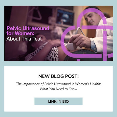
Demystifying Pelvic Ultrasound: A Comprehensive Guide


Pelvic Ultrasound Test in Women

A pelvic ultrasound, alternatively termed a pelvic scan or pelvic sonography, serves as a valuable noninvasive diagnostic technique, utilizing sound waves to generate detailed imagery of female pelvic organs. This comprehensive guide explores the intricacies of pelvic ultrasounds, shedding light on their purpose, procedure, and benefits, including their diagnostic capabilities.


How Does a Pelvic Ultrasound Operate?

During a pelvic ultrasound, a skilled healthcare practitioner employs a specialized handheld device known as a transducer. This transducer emits high-frequency sound waves, which penetrate the body and return as echoes upon encountering various tissues. These echoes are captured by the transducer and translated into real-time images via a computer. These resulting images offer vital insights into the size, shape, and texture of pelvic organs.


Varieties of Pelvic Ultrasound

There are two common approaches to conducting a pelvic ultrasound:


1. Transabdominal Ultrasound: In this method, the transducer is placed on the lower abdomen, with conductive gel applied to optimize sound conduction. The transducer is moved across the abdomen to capture images, often aided by a full bladder to enhance visibility.


2. Transvaginal Ultrasound: This approach entails inserting a lubricated transducer into the vagina, affording closer proximity to pelvic organs for clearer and more detailed images. It is advisable for the patient to empty their bladder before the procedure for added comfort.


The Pelvic Ultrasound Procedure

Typically, a pelvic ultrasound is a painless and straightforward process, involving preparation, transducer placement, image capture, and, in some instances, a Doppler ultrasound to assess blood flow. The procedure's duration generally ranges from 15 to 45 minutes, contingent on specific case requirements.


What Can a Pelvic Ultrasound Discern?

Pelvic ultrasounds offer numerous benefits, including highly detailed imaging for accurate diagnosis, safety without radiation exposure (including for pregnant women), efficiency with rapid real-time results, and versatile applications across gynecology and pregnancy monitoring. These advantages contribute to timely diagnosis, effective treatment planning, and improved patient outcomes.


In summary, a pelvic ultrasound is a precise and secure method for evaluating female pelvic organs. Its provision of clear visuals, quick results, safety, and versatility renders it an invaluable diagnostic tool in gynecology and pregnancy care. For inquiries or additional information concerning the advantages of pelvic ultrasounds in diagnostics, consultation with a healthcare professional is recommended. To gain a more comprehensive understanding of pelvic ultrasounds, we invite you to read the full blog on Pelvic Ultrasound Test in Women. Your knowledge about this diagnostic procedure is just a click away.

Thursday 12 October 2023

The Impact of High Altitude on Lung Capacity and Heart Health


How High Altitude Affects the Body

High-altitude destinations, known for their majestic peaks and stunning vistas, beckon adventurers worldwide. Yet, ascending to these heights triggers significant bodily changes that profoundly impact heart and lung health. The relationship between high altitudes and their effects on the cardiovascular and respiratory systems is explored in detail in the full blog on the Trivitron Blog. 


At high altitudes, lung capacity is notably affected due to the thinning of the air, which contains fewer oxygen molecules per breath. This scarcity of oxygen compels the body to work harder to extract oxygen for cellular processes, prompting an increase in ventilation rate to maintain oxygen levels.


The challenges posed by high altitudes affect the body, especially the heart. While the heart generally adapts to altitude well, those with pre-existing heart conditions may face difficulties. High altitudes can exacerbate conditions such as hypertension, adding strain to the heart. The heart may also experience arrhythmias as it copes with heightened oxygen demands.


The heart's oxygen problem becomes apparent at high altitudes, where thin air provides fewer oxygen molecules for cellular functions. To compensate, the heart pumps more blood to deliver vital oxygen to cells, ensuring their sustenance.


In conclusion, high-altitude travel offers unparalleled beauty and tranquility, but understanding its impact on lung capacity, overall body function, and potential challenges, particularly for individuals with heart conditions, is crucial. The full blog provides a comprehensive understanding of these crucial aspects and offers tips for safely navigating high altitudes, which are paramount for your health and safety.

Source